
Modern CT and MRI scanners produce terabytes of data daily. AI systems now process these scans in seconds, flagging critical findings that human eyes might miss.
Radiologists Aren't Being Replaced — They're Being Drowned
The narrative that AI will replace radiologists is dead wrong. What's actually happening is far more interesting — and more urgent. Global imaging volumes are growing at 5-7% annually while the radiologist workforce grows at barely 2%. The United States alone performed over 3.6 billion imaging procedures in 2025. That's 11 scans per person. Meanwhile, the average radiologist reads between 100 and 200 studies per day, with some high-volume specialists pushing past 300. Fatigue-induced errors in the fourth hour of a reading shift run as high as 26% above baseline.
This is the real problem AI solves. Not replacement. Triage. The technology that started in research labs as a curiosity has become operational infrastructure in over 1,800 hospitals worldwide — and the data is now undeniable. What matters is not whether AI matches humans — it's where AI exceeds human capability, and where it still falls embarrassingly short.
The JAMA Study That Changed Everything
A landmark 2025 study published in JAMA evaluated five FDA-approved AI systems for mammography screening across 275,000 exams at 45 clinical sites. The results landed like a bombshell. The top-performing model matched the 87% sensitivity of double-reading radiologists — two radiologists reading the same mammogram — while simultaneously reducing false positives by 23%. In women with dense breast tissue, where conventional mammography misses roughly 50% of cancers, the AI detected 8% more malignancies. Dense breast tissue affects 40-50% of women screened, making this arguably the single most impactful demographic gain in breast imaging this decade.
The study also exposed a brutal truth: the variation in performance across different AI systems was enormous. The worst-performing system in the study hit only 74% sensitivity — 13 points behind the leader. This underscores a critical point that administrators often miss: all AI is not created equal, and buying the wrong system can be worse than buying none.
Google Health's Mammography Work: The Unfinished Revolution
Google Health's mammography AI, first published in Nature in 2020, has continued to evolve through iterative clinical deployments. Their latest model, deployed across multiple UK National Health Service sites and partner hospitals in the US, uses an ensemble architecture combining five specialized deep neural networks — each trained on different mammographic views and tissue densities. The ensemble approach reduced the false-positive rate by an additional 5.8% compared to single-model systems while maintaining sensitivity above 90%. The system has now processed over 1.2 million screening mammograms in clinical settings. A retrospective analysis from the partnership with Northwestern Medicine showed a 37% reduction in unnecessary biopsies when the AI was used as a second reader — representing not just clinical improvement but massive cost savings, given that the average diagnostic breast biopsy in the US costs between $750 and $1,500.
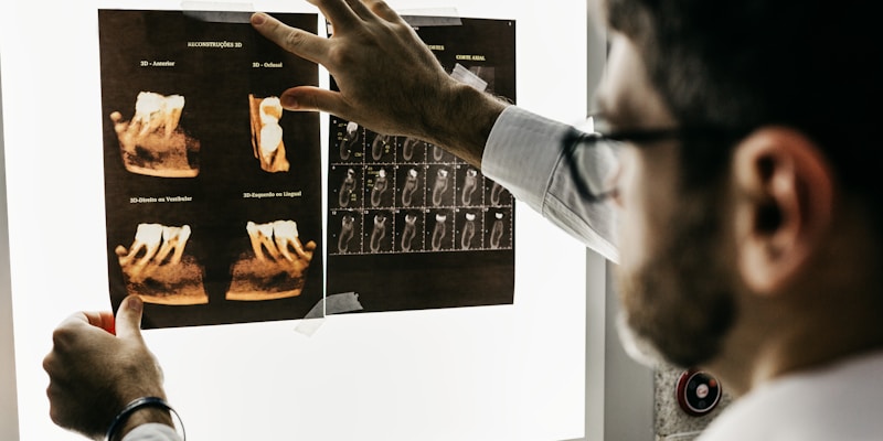
Radiologists review AI-triaged studies at a reading station. Workflow redesign is proving harder than the AI itself.
Who's Actually Winning in AI Imaging?
The competitive landscape in AI medical imaging has matured rapidly. Not all players are equal — and the data reveals a clear hierarchy. Here's how the major deployed systems stack up based on peer-reviewed outcomes through early 2026:
Aidoc: The Quiet Behemoth of Medical AI Triage
Aidoc has quietly become the most widely deployed AI imaging company on the planet, with its software now processing over 2 million radiology studies every month across 1,700 hospitals. That's roughly one study every 1.3 seconds, 24/7. The company holds FDA clearance for 14 separate clinical indications — more than any competitor — covering everything from intracranial hemorrhage detection in non-contrast head CTs to pulmonary embolism on CT angiography and incidental pulmonary nodules on chest X-rays.
The real-world numbers are staggering. At HCA Healthcare, which operates 186 hospitals across the United States, Aidoc's AI has been integrated into the PACS workflow since 2023. A retrospective analysis of 450,000 studies showed that AI-prioritized exams with critical findings were opened by radiologists an average of 32 minutes faster than non-prioritized studies. For intracranial hemorrhage — a condition where every minute counts — the median time from scan acquisition to radiologist review dropped from 56 minutes to 11 minutes. Hospitals using Aidoc's triage system reported a 41% reduction in time-to-treatment for acute stroke patients.
PathAI: Deep Learning Meets the Microscope
Pathology is arguably the area where AI has the most upside — and the most resistance. A single pathologist reviewing a biopsy slide spends 30-60% of their time on purely mechanical tasks: scanning normal tissue to find the few abnormal cells that matter. PathAI's deep learning models, trained on over 500,000 digitized slides from 300+ pathology labs, tackle exactly this bottleneck. Their models for breast and prostate cancer detection have demonstrated 99.3% sensitivity in detecting malignant regions on whole-slide images, reducing the risk of micro-metastases going undetected.
But the company's most impressive metric isn't accuracy — it's speed. At the 2025 USCAP conference, PathAI presented data showing that their AI-assisted workflow reduced pathologist slide review time by an average of 43% for breast cancer cases while maintaining diagnostic concordance with conventional review at 98.7%. In practical terms, a pathologist who previously reviewed 40 breast cancer cases per day could handle closer to 70 with AI assistance. Given the projected shortage of 5,700 pathologists in the US by 2030, that kind of throughput gain isn't nice-to-have — it's survival.
Viz.ai: Minutes Matter in Stroke Care
Viz.ai has carved out a specific and brutally important niche: stroke imaging. The company's platform automatically analyzes CT perfusion and CTA scans the moment they're acquired, identifying large vessel occlusion (LVO) strokes — the most severe type — and sending alerts directly to neuro-interventionalists' smartphones. No waiting for radiology to dictate a report. No PACS queue delays.
The NHS England deployment across 24 stroke units demonstrates why this matters. The system identified LVOs with 96% sensitivity and 93% specificity, processing scans within 60 seconds of acquisition. The clinical impact: a 14% improvement in functional independence at 90 days post-stroke, measured by the modified Rankin Scale. For every 100 patients processed through Viz.ai's system compared to standard workflow, an additional 14 patients walked out of the hospital functionally independent. The number needed to treat to prevent one disability — previously around 8 for standard mechanical thrombectomy — dropped to 5.5 with AI-assisted workflow.
Beyond stroke, Viz.ai has expanded into aortic aneurysm detection and coronary calcium scoring, processing over 1 million studies monthly across 1,200+ hospitals in the US and Europe. The company's 2025 data shows that their AI identified incidental aortic aneurysms — often silent and deadly — in 1.2% of all chest CT scans reviewed, with 97% of those patients having no prior diagnosis.
Butterfly Network: AI That Put Ultrasound in 50,000 Pockets
Butterfly Network took a radically different approach: instead of building AI for expensive hospital CT and MRI scanners, they built a $2,000 handheld ultrasound probe that connects to a smartphone, then layered AI guidance on top. The Butterfly iQ+ device has been deployed in over 50,000 clinical settings, from Harvard teaching hospitals to rural clinics in sub-Saharan Africa. The company's AI provides real-time guidance for probe positioning (eliminating the steepest learning curve in ultrasound), automatic organ recognition, and quantitative measurements.
The most telling data point comes from a 2025 study in JAMA Internal Medicine: non-expert clinicians — nurses and physician assistants with less than 2 hours of training — achieved 92% diagnostic accuracy for focused assessment with sonography in trauma (FAST) exams using the Butterfly AI system, compared to 65% without AI guidance. In emergency medicine, where minutes determine survival, turning a nurse into a passable sonographer in two hours is not incremental improvement — it's a paradigm shift.
Handheld ultrasound devices paired with AI guidance are democratizing imaging access worldwide.
Infervision: Screening China at Scale
China's Infervision has processed over 1.5 million lung cancer CT screenings, making it the most deployed AI imaging system for lung cancer in the world. The scale is frankly unmatched elsewhere. In China, where lung cancer rates are the highest globally — 715,000 new cases annually — and radiologist density in rural provinces approaches one per 100,000 people, AI is not a luxury. It's the only viable screening strategy.
Infervision's deep learning model detects pulmonary nodules with 94% sensitivity and reduces the rate of missed nodules by 12% compared to unaided radiologist reading. The system has been deployed across 500+ hospitals in 30 provinces, including mobile screening trucks that bring low-dose CT to remote villages. A 2025 study from Peking University tracked 120,000 patients screened through Infervision's mobile program and found a 22% higher early-stage detection rate (stage I lung cancer) compared to standard referral-based screening in the same regions — translating directly to better survival odds, given that 5-year survival for stage I lung cancer is 68% versus under 10% for stage IV.
800 FDA-Cleared Devices: A Flood or a Filter?
The FDA has now cleared over 800 AI-enabled medical devices, with radiology accounting for 75% of that total. But clearance numbers are a misleading benchmark. Of those 800 cleared devices, fewer than 200 have published real-world validation data. Fewer than 100 have been adopted by more than 50 hospitals. The vast majority of cleared AI imaging products remain in pilot purgatory — used in a handful of sites, producing no meaningful outcomes data.
The FDA's updated 2025 guidance attempts to close this gap, requiring all high-risk AI devices to submit real-world performance monitoring data through mandated 12-month post-market surveillance studies incorporating diverse patient populations. The guidance explicitly addresses a problem the agency has been criticized for: AI systems that work well in neat academic datasets often degrade by 10-15% when deployed across different hospitals, scanner manufacturers, and patient demographics. A 2025 analysis of 36 cleared AI chest X-ray systems found that performance dropped an average of 8.4% when tested on external data — and 12 systems dropped by more than 20%. That's not a marginal issue. That's a systemic vulnerability.
The $500,000 Problem
The biggest barrier to AI adoption isn't the technology — it's the integration cost. A 2025 survey published in Radiology found that 62% of radiologists report using AI tools, but only 28% report significant workflow improvements. The gap between having an AI tool and it actually improving your workflow is bridged by some very expensive infrastructure.
A typical hospital PACS integration costs between $200,000 and $500,000 per AI application. For a hospital that wants to deploy AI for chest X-rays, CT head exams for hemorrhage, pulmonary embolism studies, and mammography — four of the most common applications — the total integration bill hits $800,000 to $2 million before accounting for annual maintenance and cloud compute costs. Smaller and rural hospitals are left out entirely. The economics create a two-tier system: large academic medical centers with robust IT departments adopt AI at scale, while smaller community hospitals serving disproportionately vulnerable populations continue with unaided reads. This is not a bug in the deployment model — it's a feature of a marketplace that prioritizes hospital systems with the deepest pockets.
Alert fatigue is the second-biggy problem hiding in plain sight. A 2025 analysis at a large Midwestern health system found that clinicians received an average of 41 AI-generated alerts per 8-hour shift across the five AI systems deployed. Of those, 63% were false positives. Over time, radiologists started ignoring alerts — exactly the opposite of what the technology is supposed to achieve. The lesson: deploying AI without redesigning the notification workflow is worse than deploying nothing at all.
The Multimodal Frontier: Where Imaging Meets Everything Else
The most exciting work happening right now isn't in imaging alone — it's in multimodal AI that fuses imaging data with electronic health records, genomics, and lab results. Stanford's 2025 research on multimodal breast cancer risk prediction is the current gold standard. Their model combines mammographic images with clinical risk factors — age, family history, BRCA mutation status, hormonal therapy history — to predict 5-year breast cancer risk. The model achieves an AUC of 0.89, compared to 0.75 for the Tyrer-Cuzick risk model that remains the clinical standard. That 0.14 AUC difference translates to 18% better discrimination: in a cohort of 100,000 women, the multimodal model correctly reclassifies 1,800 women who would go on to develop cancer within 5 years from "low risk" to "high risk," enabling earlier intervention.
Mass General Brigham's multimodal sepsis prediction system takes a different approach: it combines chest X-ray findings with vitals, lab values, and medication administration data to predict sepsis onset 4-6 hours earlier than standard clinical scoring. In a 2025 prospective study across five hospitals, the system reduced sepsis mortality by 12.6% and reduced ICU length of stay by 1.8 days. These are not academic curiosities — they are operational systems saving lives and money right now.
The Bottom Line: What Real Radiologists Think
The most honest assessment comes from practicing radiologists, not from company press releases or academic papers. A 2025 survey of 1,200 radiologists in the American College of Radiology's annual workforce report found that 68% believe AI will significantly change their specialty within 5 years. But only 22% believe it will reduce the demand for radiologists. The majority — 61% — believe AI will increase their value by enabling them to focus on complex cases and reduce the cognitive load of screening normal studies.
The truth is probably somewhere in between. AI is not replacing radiologists. But AI is absolutely replacing radiologists who don't use AI — a version of the famous Andrew McAfee observation that has now been validated across diagnostic imaging. The hospitals with the best clinical outcomes in 2026 are not the ones with the fanciest scanners or the most experienced radiologists. They are the ones that have figured out how to integrate AI into a well-designed workflow without drowning their clinicians in false alarms. That's the real challenge — and it's a software and operations problem, not an AI problem.